Small World Photomicrography Winners Showcase The Beauty Of The Tiny
This article is more than 2 years old
 We marvel at the wonders of our universe on a daily basis here at GFR, but if we’re being honest then that usually means drooling over some new batch of astronomy pictures. But science is forever revealing that there is just as much beauty in the very, very small as there is in the heavens above. For proof, you need look no further than the 2013 winners of Nikon’s Small World Photomicrography Competition.
As you might guess from that moniker, the competition challenges photographers to serve up images of the microscopic world all around us, and a whole different reality that usually escapes our notice because it’s too small to be seen with the naked eye. Nikon posted 20 winners for this year, and you can see the gorgeous results throughout this post.
Up top you can see the first place winner, a “Chaetoceros debilis (marine diatom), a colonial plankton organism (250x)” photographed by Wim van Egmond of Rotterdam, the Netherlands.
And here’s the second place winner: “Chrysemys picta (painted turtle) retina (400x),” photographed by Dr. Joseph Corbo of St. Louis, Missouri.

Third place: “Marine worm (20x),” photographed by Dr. Alvaro Migotto of Sao Paulo, Brazil

Fourth place: “Paramecium sp. showing the nucleus, mouth and water expulsion vacuoles (40x),” photographed by Rogelio Moreno Gill of Panama City, Panama

Fifth place: “Hippocampal neuron receiving excitatory contacts (63x),” photographed by Dr. Kieran Boyle of Glasgow, Scotland

Sixth place: “Chamaeleo calyptratus (veiled chameleon) embryo showing cartilage (blue) and bone (red),” photographed by Dorit Hockman of Cambridge, United Kingdom

Seventh place: “Adhesive pad on a foreleg of a ladybird beetle (Coccinella septempunctata) (20x),” photographed by Dr. Jan Michels of Kiel, Alberta, Germany

Eighth place: “Barbilophozia sp. (a leafy liverwort, bryophyte plant) and cyanobacteria (50x),” photographed by Magdalena Turzanñska of Wroclaw, Poland

Ninth place: “Insect wrapped in spider web (85x),” photographed by Mark A. Sanders of Minneapolis, Minnesota

10th place: “Thin section of a dinosaur bone preserved in clear agate (10x),” photographed by Ted Kinsman of Rochester, New York

11th place: “Macrobrachium shrimp (ghost shrimp) eye (140x),” photographed by Vitoria Tobias Santos of Macaé, Rio de Janeiro, Brazil

12th place: “Silicon dioxide on polydimethylglutarimide-based resist (200x),” photographed by Dr. Pedro Barrios-Perez of Ottowa, Ontario, Canada
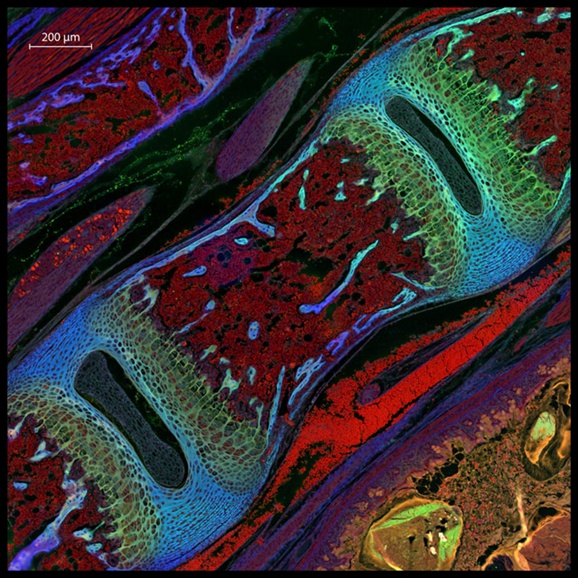
13th place: “Mouse vertebra section (200x),” photographed by Dr. Michael Paul Nelson & Samantha Smith of Birmingham, Alabama
14th place: “Peripheral nerves in E11.5 mouse embryo (5x),” photographed by Zhong Hua of Baltimore, Maryland

15th place: “Podospora anserina (fungus) filamentous tip cells (630x),” photographed by Dr. Christian Q. Scheckhubler of Frankfurt, Germany

16th place: “Pityohyphantes phrygianus (sheet weaver spider) with a parasitic wasp larva on the abdomen (5x),” photographed by Geir Drange of Asker, Norway

17th place: “Pyramidal neurons and their dendrites visualized in the visual cortex of a mouse brain (40x),” photographed by Dr. Alexandre William Moreau of London, United Kingdom

18th place: “Annelid larva (100x),” photographed by Christian Sardet of Strasbourg, France

19th place: “Nerve and muscle thin section (40x),” photographed by Dr. David Ward of Oakdale, California

20th place: “The explosive dynamics of sugar transport in fat cells,” photographed by Dr. James Burchfield of Sydney, New South Wales, Australia

You can see the runners up and more information about the winners at the Small World Photomicrography competition website.